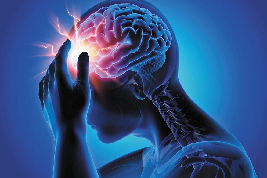
Imagine you’re in the ER, and a patient is rushed in with a suspected traumatic brain injury (TBI). Time is critical, and rapid diagnosis is essential. But where do you start? You turn to the neuro exam, one of the most fundamental yet powerful neurological tools.
Within this examination, the pupillary response is a valuable indicator, providing a glimpse into the severity of the trauma inflicted on the brain. Understanding the pupillary response in traumatic brain injury could enhance your ability to diagnose, prognose, and ultimately optimize patient care.
Understanding Pupillary Response
The pupillary response is a reflex that controls the diameter of the pupil. This seemingly simple mechanism, however, results from a complex interplay of neural pathways regulated by the autonomic nervous system.
The autonomic nervous system can be divided into two main components: the sympathetic and parasympathetic systems. These systems usually work together, each counteracting the other, thus maintaining a delicate balance. The sympathetic system, often called the “fight or flight” system, causes pupils to dilate, a state known as mydriasis.
On the other hand, the parasympathetic system, associated with “rest and digest” functions, causes the pupils to constrict. In healthy individuals, pupillary responses can be easily observed. When exposed to bright light, the pupils constrict as the parasympathetic system activates to reduce light exposure to the retina. This is called the pupillary light reflex. Conversely, in dim light or during cognitive effort, the pupils dilate as the sympathetic system kicks in to allow more light into the eye and enhance vision.
Pupillary Response in Traumatic Brain Injury
Traumatic brain injury disrupts this equilibrium, leading to alterations in pupillary response. This change could be due to direct damage to the neural pathways controlling the response or secondary to increased intracranial pressure affecting the brainstem. Understanding this deviation from normal pupil response offers an objective measure of TBI severity, complementing the overall clinical picture.
Quantifying Pupillary Response
The traditional way of pupil measurement involves a penlight and the clinician’s observation—a subjective method. Enter advanced technology, like pupillometers, which provide an objective, reproducible method for quantifying pupillary response. This device measures the neurological pupil index (NPi), an automated calculation of pupil reactivity, adding a new dimension to the neuro exam, particularly in TBI assessment.
Pupillary Response and TBI Diagnosis
A pupillary examination is a valuable part of the initial assessment and diagnosis of TBI. It complements other diagnostic methods, such as CT scans, by providing real-time bedside information about the patient’s neurological status. Abnormalities in pupillary response, such as anisocoria or a sluggish reaction to light, can be red flags, prompting immediate intervention and potentially serving as a screening tool in emergency departments.
Pupillary Response as a Prognostic Indicator
The pupillary response also carries prognostic value in TBI cases. For instance, a persistently unreactive pupil or a significant difference in the size of the pupils could signify severe brain damage and a worse prognosis. Consequently, monitoring pupillary responses could guide critical treatment decisions and help tailor patient management strategies.
Limitations and Challenges
However, interpreting pupillary responses in TBI is challenging. Factors such as the use of certain medications or the presence of pre-existing conditions can influence pupillary responses. Therefore, while assessing pupillary response is important, it must be considered in conjunction with other clinical indicators for a comprehensive assessment of TBI.
Advances in Pupillometry Research
Research in pupillometry continues to evolve, with ongoing studies exploring new frontiers. Researchers are harnessing the potential of pupil response to further our understanding of TBI management and prognosis. Integrating pupillometry data with artificial intelligence and machine learning algorithms presents an exciting avenue for future advancements in this field.
Conclusion:
The importance of pupillary responses in traumatic brain injury cannot be overstated. It is vital to the neurologic exam, providing valuable diagnostic and prognostic insights. While it’s not without challenges, technological advancements, and ongoing research promise to further enhance our understanding and capabilities in this area.
As medical professionals, staying informed and collaborating to refine our methods for diagnosing, treating, and ultimately improving patient outcomes in traumatic brain injury cases is crucial. By harnessing the full potential of pupillary response and other neurological tools, we can continue to make strides in providing the best possible care for those affected by TBI.